富山大学 システム機能形態学研究室 Systems Function and Morphology Lab., University of Toyama

音を認知する神経回路の構造を研究しています。 We study the neuronal circuitry for sensing sounds.

第4回 富山大学脳科学交流会 を行いました。 The fourth Annual Meeting of Neuroscience in Toyama

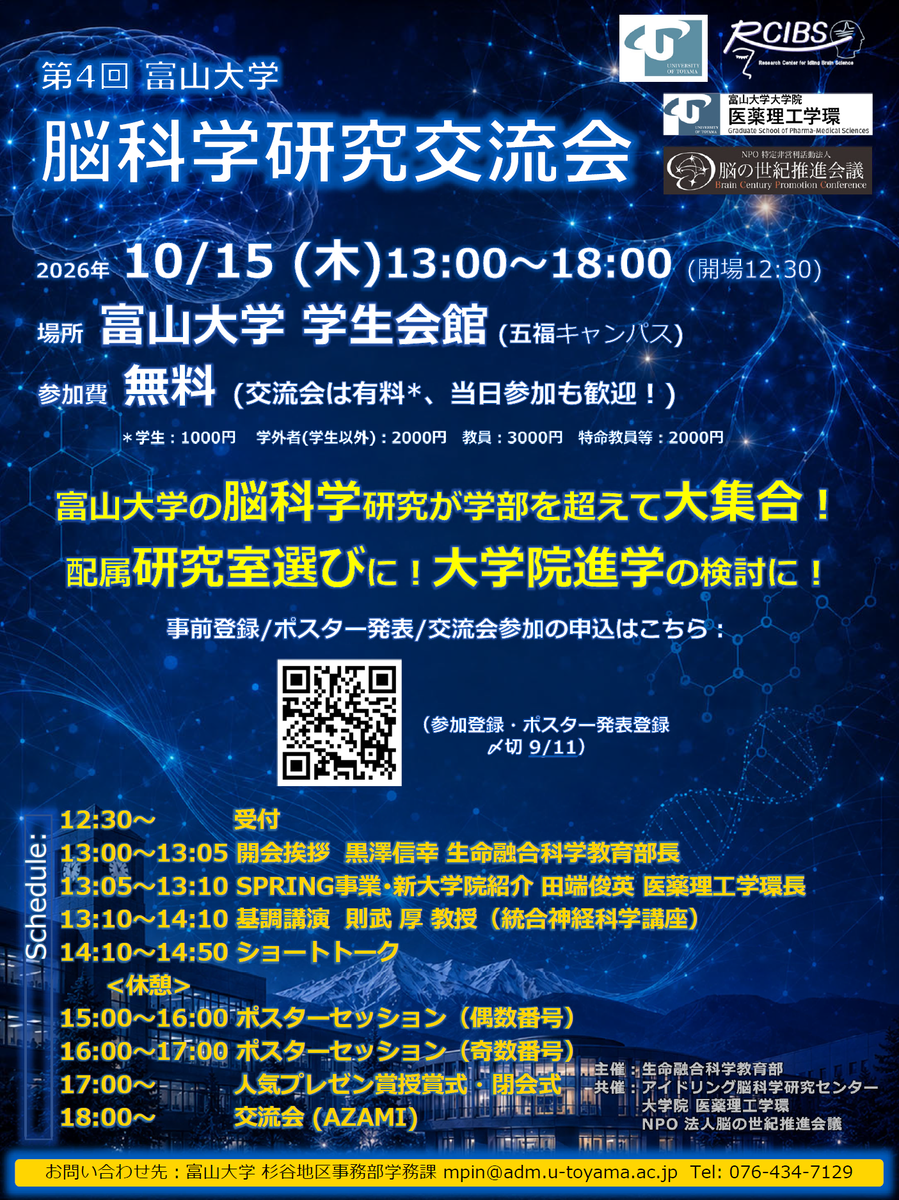
2026年10/15(木)に富山大学脳科学交流会を行いました。これで4回目となります。


今回、基調講演を担当したのはこの4月に統合神経科学講座教授に着任された則武厚先生で、「Beyond Reward: Neural Mechanisms of Social Decision-Making and Emotions in Primates」という演題で講演してもらいました。同じ報酬であっても周りの仲間との比較で報酬の価値が変化してしまう、というよくある経験がどのような脳内メカニズムで起きるのか、という話で多くの人が大変興味深く聞いていました。

続くショートトークセッションでは11名の方が発表しましたが、今回特筆すべきこととして、他大学からの来訪者も発表したことです 。本研究室で2週間の日程で共同研究に来ていたセント・アンドリュース大学のBenjamin Thompsonさんが本研究室との共同研究の内容で発表を行ってくれました。

ポスターセッションでは50題発表がありました。今回も学生ポスターについて選考が行われ、生化学講座のHassan Rubyさんが最優秀ポスターに選ばれました。

Hassan Rubyさんに賞状を手渡す筆者

今回、優秀賞などのサポートに、NPO法人「脳の世紀推進会議」からの支援をいただきました。この場を借りてお礼申し上げます。


交流会にもたくさんの方が参加され、大盛況でした。


これまで「脳科学交流会」は大学院・生命融合科学教育部のイベントとして実施されてきました。生命融合科学教育部の学生は基本今年度で全員卒業ですので運営母体がなくなってしまいます。参加者からの意見などを踏まえて今後の実施について検討していきたいと思います(基本的にはやりたいと思っております)。

論文「The Nucleus of the Brachium of the Inferior Colliculus Contains Thalamic-Like and Midbrain-Like Subregions(下丘腕核には視床様領域と中脳様領域を含む)」の公開 Our paper is now available online!

伊藤が筆頭著者の論文、The Nucleus of the Brachium of the Inferior Colliculus Contains Thalamic-Like and Midbrain-Like Subregionsが Journal of Comparative Neurology誌にて公開されました。

Ito’s first-author paper, “The Nucleus of the Brachium of the Inferior Colliculus Contains Thalamic-Like and Midbrain-Like Subregions”, has been published in the Journal of Comparative Neurology.


下丘腕核はその名前からわかるように、下丘という中脳の脳領域の一部と考えられてきました。下丘は聴覚神経回路のメイン領域の一つで、聴覚情報を脳幹の様々な領域から受けて、視床にその情報を送ります。視床が大脳皮質に情報を伝えることで知覚が生じると考えられています。重要なのは、大脳皮質に向かう感覚情報は必ず視床で中継されるということです。

As its name suggests, the nucleus of the brachium of the inferior colliculus (nBIC) has traditionally been regarded as part of the inferior colliculus, a midbrain structure. The inferior colliculus is a major hub in the auditory system, receiving auditory information from various brainstem regions and relaying it to the thalamus. The thalamus then transmits this information to the cerebral cortex, where perception is thought to arise. A key principle is that sensory information reaching the cerebral cortex must be relayed by the thalamus.


ところが、我々の2025年の論文で、下丘腕核から大脳皮質への直接投射があることが明らかになりました。その直後に海外のグループでも同様の報告がありました。このことは下丘腕核が視床としての性質をもつことを示唆しています。一方、別の論文は下丘腕核が下丘と同様の細胞構築をもつことを示しており、こちらは下丘腕核が中脳の性質をもつことを示唆しています。中脳と視床は発生学的にも異なる構造であり、両方の性質を持つ、ということはなかなか考えがたいことです。本研究は神経回路解析や遺伝子発現解析を行うことで、下丘腕核の前部に大脳皮質に投射する細胞群が、後部には視床に投射する細胞群があり、両者は分離していることを明らかにしました。

However, in our 2025 paper, we demonstrated a direct projection from the nBIC to the cerebral cortex. Shortly afterward, an overseas research group reported a similar finding. These observations suggest that the nBIC has thalamic-like properties. Meanwhile, another study showed that the nBIC has a cellular architecture similar to that of the inferior colliculus, suggesting midbrain-like properties. Because the midbrain and thalamus are developmentally distinct structures, it is difficult to reconcile these two characteristics within a single nucleus. In the present study, neural circuit mapping and gene expression analyses revealed two separate populations: neurons projecting to the cerebral cortex in the anterior nBIC and neurons projecting to the thalamus in the posterior nBIC.


大脳皮質に投射するものも、視床に投射するものも、視覚、聴覚、体性感覚情報を受け取ることがわかっています。このことから、下丘腕核は多感覚情報を統合する脳領域で、それを大脳皮質に直接送るものと、皮質外に送るという情報の仕分けを行っていると推測されます。

Both the cortex-projecting and thalamus-projecting populations are known to receive visual, auditory, and somatosensory information. This suggests that the nBIC integrates information across sensory modalities and routes it either directly to the cerebral cortex or to subcortical structures.


下丘腕核は、大脳皮質へ広範な脳領域に散在性の出力を送るほか、その途上で尾側線条体という感覚に関係する大脳基底核へも側枝をつくります。大脳皮質への出力は極めて潜時が短いことから、音に対して幅広い大脳皮質領域を活性化する働きがあると考えられます。線条体は習慣的な情報を学習すると考えられており、線条体への出力は学習された感覚情報の想起に関係するのかもしれません。

The nBIC sends diffuse projections to widely distributed cortical regions. Along the way, these projections also give off branches to the caudal striatum, a sensory-related part of the basal ganglia. The very short latency of its cortical output suggests that the nBIC helps activate widespread cortical regions in response to sound. The striatum is thought to support habit learning, and the nBIC’s striatal output may contribute to the recall of learned sensory information.


下丘腕核の皮質外への出力として、視床の他に重要なものが上丘です。上丘は視覚などの感覚刺激を受けてその方向に視線を向けるのに重要な働きを持ちます。下丘腕核から上丘への投射は音の空間情報を伝えると考えられていて、我々の最近の論文では上丘における視覚と聴覚の相互作用の基盤となる構造が見出されています。

In addition to the thalamus, another important subcortical target of the nBIC is the superior colliculus. The superior colliculus plays a key role in directing gaze toward visual and other sensory stimuli. The projection from the nBIC to the superior colliculus is thought to convey information about sound location. In our recent paper, we identified an anatomical substrate for interactions between vision and hearing in the superior colliculus.


このように皮質への出力と皮質外への出力には機能的な違いがあるように思われます。本研究で見出された神経回路がこのような機能的仕分けを行っていると考えています。

Thus, the cortical and subcortical outputs of the nBIC appear to serve different functions. We propose that the neural circuitry identified in this study supports this functional routing of sensory information.


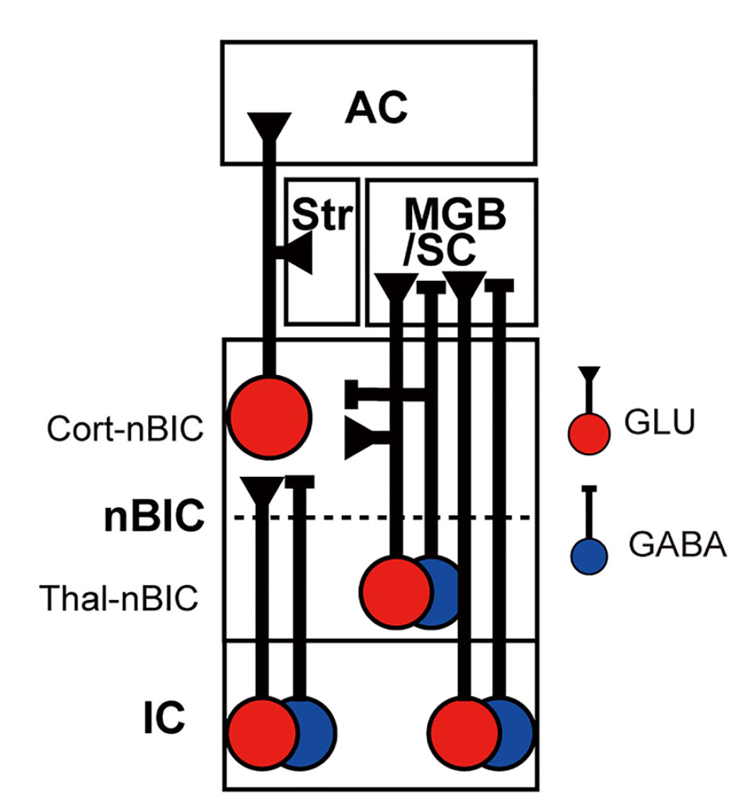
回路の概念図:下丘(IC)には興奮性(GLU)、抑制性(GABA)の2種類の細胞がおり、一方下丘腕核(nBIC)は前部(Cort-nBIC)は興奮性のみ、後部(Thal-nBIC)は興奮性と抑制性の両方を含む。後部は視床(MGB)と上丘(SC)に投射し、前部は聴覚野(AC)と線条体(Str)に投射する。

Schematic of the neural circuitry: The inferior colliculus (IC) contains both excitatory glutamatergic (GLU) and inhibitory GABAergic (GABA) neurons. In the nBIC, the anterior subdivision (Cort-nBIC) contains only excitatory neurons, whereas the posterior subdivision (Thal-nBIC) contains both excitatory and inhibitory neurons. The posterior subdivision projects to the medial geniculate body of the thalamus (MGB) and the superior colliculus (SC), while the anterior subdivision projects to the auditory cortex (AC) and striatum (Str).


さくらサイエンスプログラムの活動を終えて Reflecting on the Completion of the Sakura Science Exchange Program

2026年8月23日から9月5日までの2週間、JSTさくらサイエンスプログラムの支援を受け、国立台湾大学(NTU)生命科学院の呂昕煒(Hsin-Wei Lu)助教と学生4名を研究室に迎えました。呂博士とは2025年夏の日本神経科学学会で知り合い、共通する聴覚神経回路研究を通じて共同研究の機会を探ってきました。その後、本学とNTU生命科学院との学術交流協定も締結され、今回の招聘は交流を具体化する絶好の機会となりました。

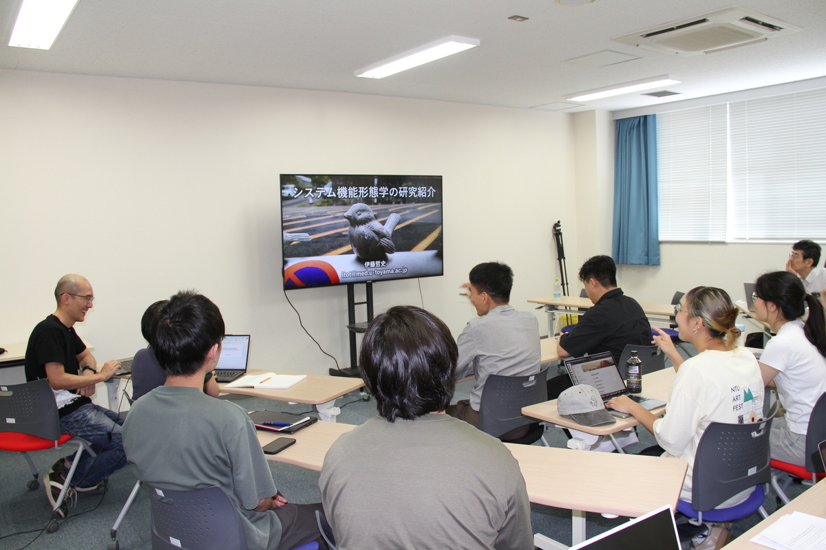
受け入れに当たっては、本学国際課の支援のもと、動物実験・遺伝子組換え実験に必要な申請や事前教育を完了し、到着後すぐに研究活動を開始できる体制を整えました。初週には、双方のスタッフと学生が英語で研究発表を行い、研究内容や実験技術への理解を深めました。これは本研究室の学生にとっても、英語による発表と議論を経験する貴重な機会となりました。

活動前半は研究・実験の見学、後半は実践を中心としました。NTUの学生3名は、本研究室独自の脳定位手術法を熱心に練習し、納得できるまで粘り強く実験を続けました。博士課程のYao-Hsinさんは、研究室にあった装置を活用して、難易度の高いin vivo whole-cell記録系の立ち上げに挑戦しました。対象脳領域では報告例が極めて少ない手法ですが、限られた滞在期間中に見事成功し、興味深い記録を得ました。この成果は本研究室のスタッフにも大きな刺激を与えました。

実験の合間には、神経科学研究を行う学内5研究室を訪問しました。睡眠脳ダイナミクス研究室の大学院生の宮本さんは、中国語に堪能で、NTU神経科学サマースクールへの参加を通じて一行とすでに交流があり、滞在中の生活や文化体験も支援してくれました。休日には岩瀬の歴史的町並みを巡り、「おわら風の盆」も訪れるなど、富山の文化に触れる機会を設けることができました。

9月4日の最終報告会では、各学生が研修成果を発表しました。続いて、呂博士が本学総合医薬学研究科の特別大学院セミナーで講演し、医学部・理学部の教員や学生、国際機構関係者らと活発な議論を行いました。懇親会では、NTUとのさらなる交流や学生支援、共同研究の発展に向けた具体的な案も話し合われました。

NTU学生(左4名)の最終プレゼンテーション。伊藤(右上)、呂(右下)

Final presentation by NTU students (left). Discussing with Ito (right-top) and Lu (right).

大学院セミナー。謝辞のスライドで、さくらサイエンスプログラムへの言及。

Graduate school seminar. This is the acknowledgement slide with reference to Sakura Science Program.

懇親会にて。大学のトートバッグに参加者全員のサイン

At socials. Signatures of all attendees on University tote bags.


今回の招聘は、一方向の技術紹介にとどまらず、双方が刺激を受け、研究水準を高め、新たな共同研究を生み出す契機となりました。呂博士との共同研究やYao-Shinさんへの研究指導は今後も継続する予定です。この成果を本学とNTUのより高度な連携へと発展させていきたいと考えています。貴重な機会を与えてくださったJSTさくらサイエンスプログラムの皆様に、心より感謝申し上げます。


From August 23 to September 5, 2026, our laboratory hosted Assistant Professor Hsin-Wei Lu and four students from the Department of Life Science, College of Life Science at National Taiwan University (NTU), with support from the JST Sakura Science Exchange Program.


Dr. Lu and I first met at the Annual Meeting of the Japan Neuroscience Society in the summer of 2025. We quickly discovered our shared interest in auditory neural circuits and began exploring opportunities for collaboration. Earlier this year, I participated in a joint seminar for faculty members from NTU and our university and was deeply impressed by the high caliber of NTU’s students. Subsequently, with the support of our university’s international affairs organization, an academic exchange agreement was concluded between NTU’s College of Life Science and our university. The Sakura Science program therefore provided a timely opportunity to turn this growing relationship into concrete research collaboration.


With extensive assistance from our International Affairs Division, we completed all necessary preparations before the visitors arrived. Because our research involves animal experiments and genetically modified organisms, every participant had to be included in approved research protocols. Advance training enabled the students to receive authorization smoothly and begin laboratory work immediately.


During the first week, members of both laboratories gave short research presentations in English. These sessions allowed the NTU visitors to understand our research projects and experimental techniques while giving our students valuable experience presenting and discussing science internationally.


The first half of the program focused mainly on observing our laboratory’s research activities, while the second half emphasized hands-on experiments. Three NTU students practiced our stereotaxic surgery procedures, which differ from those used in their laboratory. Their perseverance was remarkable: they continued working until they were fully satisfied with their results, sometimes remaining in the laboratory until after the last bus had departed.


Yao-Hsin, a doctoral student, noticed a patch-clamp amplifier and pipette puller in our laboratory and proposed establishing an in vivo whole-cell recording system. This is an extremely challenging technique, with only a few published studies involving the brain region we investigate. Nevertheless, within the limited period available, Yao-Hsin successfully established the procedure and obtained highly interesting recordings. His achievement greatly inspired our laboratory members, particularly Assistant Professor Hase, who conducts our physiological experiments and became eager to master the technique himself.


As the University of Toyama’s Faculty of Medicine has many laboratories engaged in neuroscience research, the visitors toured five laboratories between experiments. Ms. Miyamoto, a graduate student in the Sleep and Brain Dynamics Laboratory, had already met the visitors through the NTU Neuroscience Summer School and provided invaluable support throughout their stay. This demonstrated that exchanges with NTU were already developing into a broader academic network.


Although the participants spent most days conducting experiments from morning until evening, they also experienced Toyama’s culture. They explored the historic streets of Iwase after traveling there by canal boat and attended the famous Owara Kaze-no-Bon festival, where dancers in traditional hats perform to the evocative sound of the kokyu.


On September 4, the students presented their achievements at a final research meeting. Dr. Lu then delivered a special graduate seminar attended by faculty members and students from several departments, as well as representatives of the university’s international and global research organizations. The seminar generated lively discussion, and the following reception produced several ideas for expanding exchange and collaboration with NTU.


This visit was far more than a one-way transfer of experimental techniques. It enabled researchers and students from both institutions to learn from one another, improve their research, and establish a strong foundation for joint projects. Our collaboration with Dr. Lu’s laboratory will continue, including further research guidance for Yao-Hsin. We hope to build a sustainable framework supporting students engaged in these collaborations and to develop an even stronger partnership between our university and NTU.


We sincerely thank everyone involved in the JST Sakura Science Exchange Program for providing this valuable opportunity.

[for contacting to Tetsufumi Ito, e-mail: itot<at>med.u-toyama.ac.jp (Please replace <at> with @)]


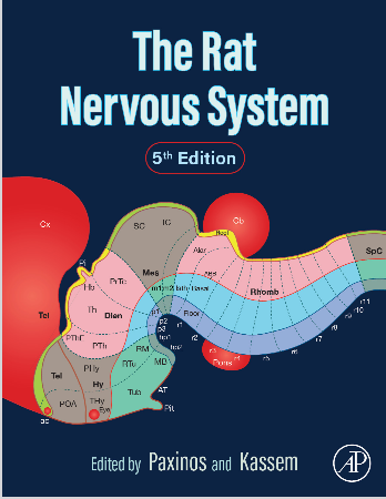
伊藤が執筆した本が出版されました!:Rat Nervous System, 5th ed is now published!

伊藤が執筆に協力したGeorge Paxinos等が編者のシリーズThe Rat Nervous Systemの第5版が出版されました! 伊藤はサラマンカ大学のManuel Malmierca教授と共著で第28章「Auditory System」を担当しました。末梢(蝸牛)から聴覚野に至る聴覚伝導路全体における形態と機能の両面をラットやマウスの研究を中心に包括的に取り上げています。

shop.elsevier.com


The fifth edition of The Rat Nervous System, edited by George Paxinos and colleagues, has just been published, and Tetsufumi Ito contributed to this new edition!

Together with Professor Manuel Malmierca of the University of Salamanca, Ito co-authored Chapter 28, “Auditory System.” The chapter provides a comprehensive overview of both the anatomy and function of the entire auditory pathway, from the periphery (cochlea) to the auditory cortex, with a particular focus on research in rats and mice.


国立台湾大学Lu博士グループ来学

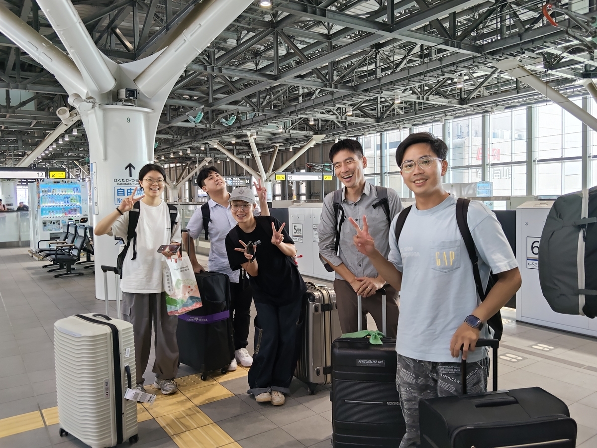
JSTさくらサイエンス事業の支援により、8/23-9/5の2週間の予定で国立台湾大学のHsin-Wei Lu博士と学生4名が本研究室に滞在して共同研究を行っています。

富山駅にて。右から2番目がLu博士。

合同セミナーの様子。皆さんとても積極的に質問していました。


本研究室の学生の皆さんにも刺激になっているみたいです。